| ON THE COVER |  |
|---|---|
| Vol. 77 No.1 March 2012 | |
| Technical note | |
|
|
|
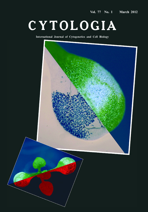
| Spontaneous Chlorotic Cell Death in atlrgB Mutants of Arabidopsis thaliana AtLrgB with 12 predictive transmembrane domains is a plastid envelope protein in Arabidopsis thaliana. Under short-day conditions, the cotyledons and true leaves of the atlrgB mutant plants show immediate greening, similar to wild-type plants, after which some parts demonstrate a chlorotic phenotype. When the atlrgB mutant is grown under continuous light, its chlorotic phenotype is suppressed. Sterilized seeds of the atlrgB mutants were planted on MS agar medium containing B5 vitamins with 2% sucrose, and grown in a cultivation room with approximately 100 μmol phoon m-2 s-1 at 23°C under 9.5-h light/14.5-h dark cycle for 2 weeks. Using a fluorescent stereo microscope (M165FC; Leica Microsystems), images of a plant on the plate and of chlorophyll autofluorescence were observed, and combined in 1 panel (left). Cotyledons of the atlrgB mutant showed chlorosis, while those in the wild-type plants stay green at 2 weeks after sowing. In the atlrgB mutant plant, first true leaves seem to develop normally. Detached cotyledon was soaked in trypan blue, a reagent for detecting cell death (75% ethanol, 8.3% TE-saturated phenol [pH 8.0], 8.3% glycerol, 8.3% lactic acid, and 0.17 g/ml trypan blue). The cotyledon was then boiled for 1 min and left overnight at room temperature. Finally, the cotyledon was washed several times with 2.5 g/ml chloral hydrate and observed under the stereo microscope (Leica). Trypan blue staining image was taken and combined with the image of the same cotyledon before staining (right panel). The degreened sectors in the cotyledon contained dead cells. In contrast, no dead cells were observed in the green sectors. The chlorotic sectors were symmetrically oriented at the margins. The detailed function of AtLrgB in plastid envelope remains unclear. Research on AtLrgB may be key to uncovering relationship between cell death and plastid function. For details, see the article by Yamaguchi et al. 2012. Plant Cell Physiol. 53: 125–134. (Mizuki Yamaguchi 1 , Katsuaki Takechi 1 and Hiroyoshi Takano 1,2 1 Graduate School of Science and Technology, Kumamoto University, Kumamoto 860–8555, Japan. 2 Bioelectrics Research Center, Kumamoto University, Kumamoto 860–8555, Japan) |
|